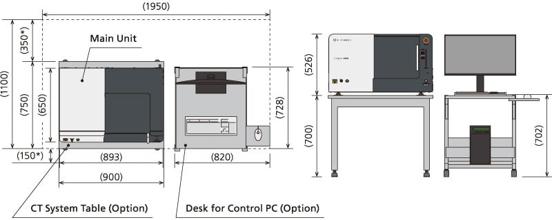



The bench-top X-ray computed tomography (CT) system XSeeker 8000 is equipped with a high-output X-ray generator and a high-resolution flat panel detector. Despite its compact size, the device has a high X-ray output voltage (160 kV), which enables inspections with high image clarity for molded plastic and metal (particularly aluminum) parts. Furthermore, the new software for the device features a high level of functionality and boasts the highest throughput in its class to date. Due to the combination of these factors, the device can be applied to solve a wide range of tasks, from detailed inspections during product development and quality evaluation to checks at production sites.
XSeeker 8000
Microfocus X-ray CT System

Features
Compact, high-performance system
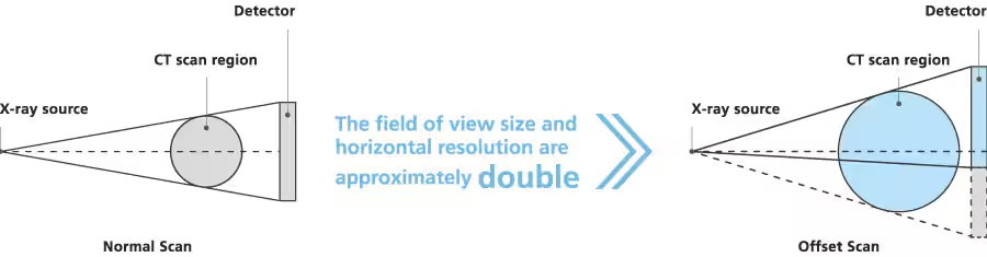
- The smallest and lightest bench-top CT system in its class.
Thanks to its compact dimensions, the device can be installed in practically any laboratory or production room.
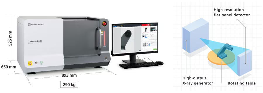
- High output voltage of 160 kV ensures high penetrability
With an X-ray output voltage of 160 kV, the system is suitable for inspecting thick parts (for example, high-density plastic or metal products)
| Material | Ratio of X-ray transmittance of other manufacturers’ models on the market to the new Shimadzu model (100 kV : 160 kV) |
|---|---|
| Plastic | 1:1.2 |
| Aluminum | 1:1.4 |
| Iron | 1:2.0 |
Typical image obtained by systems with an output voltage of 100 kV and 160 kV (transmission was performed through an aluminum plate with a thickness of 100 mm)
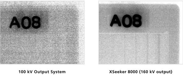
Image quality and functionality on par with high-end models
- High-precision scanning with a maximum input resolution of 5.6 million pixels.
The offset scanning technology used in the XSeeker 8000 roughly doubles the sample coverage area compared to normal scanning, allowing for a resolution equivalent to 5.6 million pixels.
The system provides high-precision scanning in a wide field of view with a diameter of up to 100 mm.
- Flat panel detector with a 50 µm pixel pitch
The system is equipped with a flat panel detector with fine image elements measuring approximately 50 µm per pixel. This is the smallest pixel pitch among all devices in this class, enabling reliable observations down to the level of fine structures.
- The base version of the device is equipped with highly functional universal software for image viewing
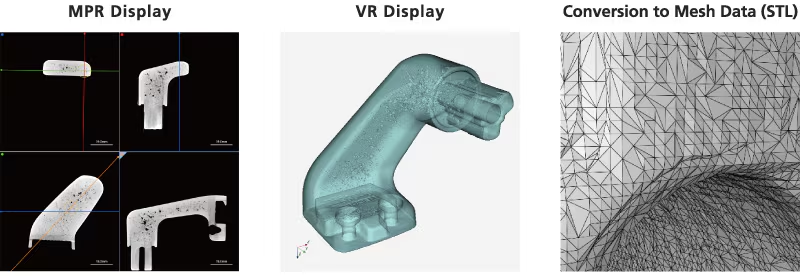
The system is equipped with specialized image viewing software featuring several operator-friendly functions. Thus, in addition to an MPR display for simultaneously displaying multiple cross-sections of the inspected object, the software includes a VR display showing a full 3D matrix of the object, allowing for inspections on an intuitive level.
The dimensional measurement function is available on both the MPR image and the VR image. The obtained CT observation results can be immediately converted into STL mesh data, significantly expanding the use of the acquired results. For example, for use in 3D CAD software or for printing on a 3D printer.
Operating mode adapted for inspection convenience
- 3-step scanning
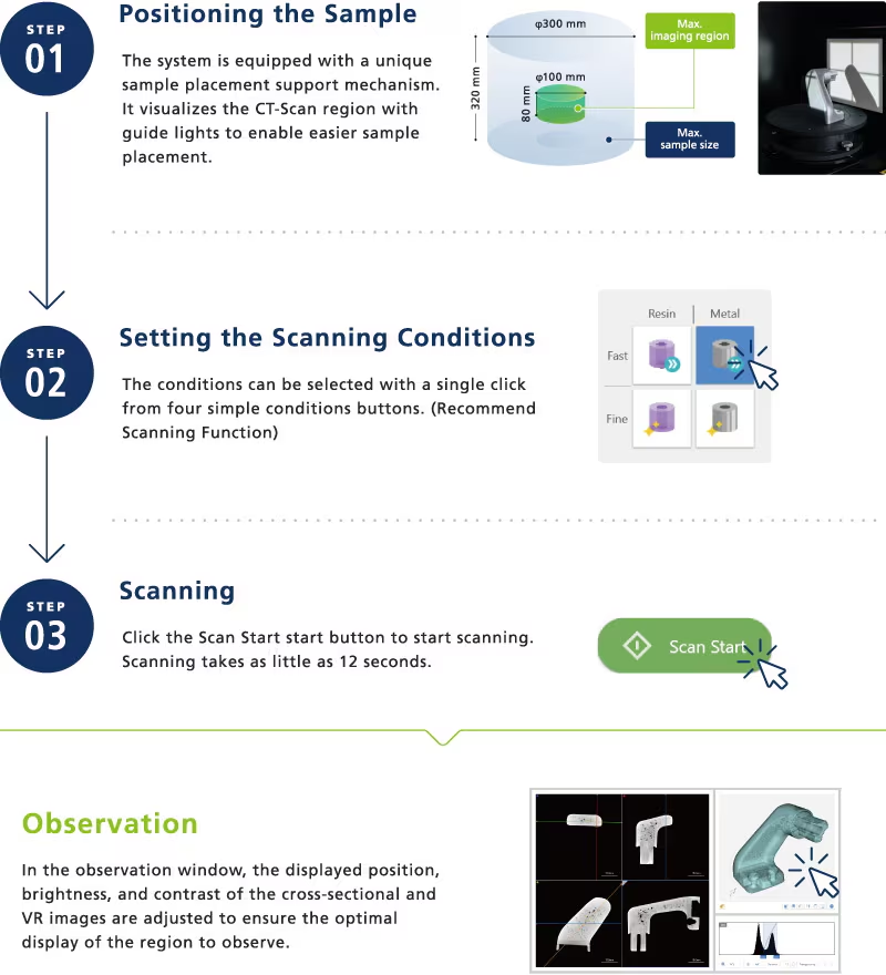
A simple three-step setup algorithm allows you to easily start scanning the inspected object. Once set up, a user with minimal knowledge can start working on the system almost immediately. The device does not require a calibration procedure before each start of operation.
- One-button operation
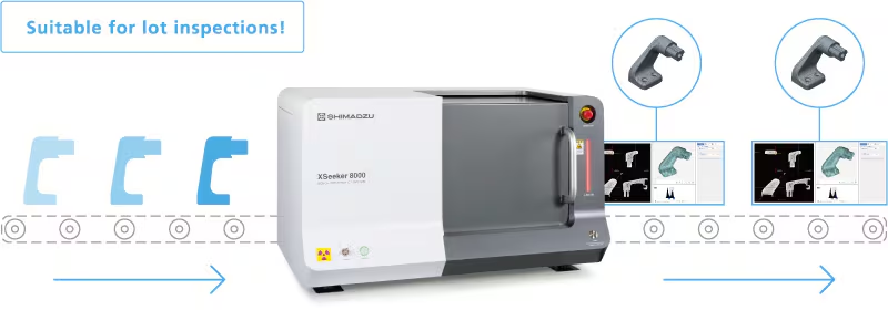
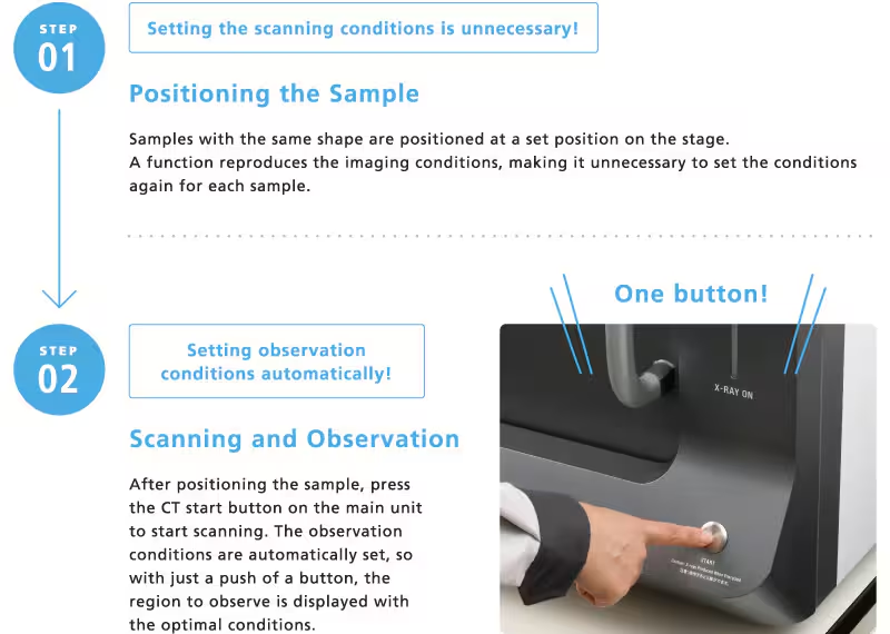
In the case of inspections involving repeated scanning of identically shaped samples, operation is carried out with the push of just one button, significantly increasing productivity.
Optimal conditions, including the displayed position and contrast during observations, are recorded along with the scanning conditions. This allows observation to begin immediately after scanning without adjusting the observation position or contrast. All operations, from starting the scan to observing the results, can be performed with the push of a single button, without any additional operations within the software.
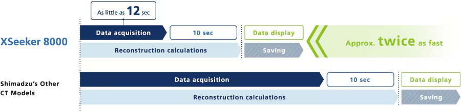
- Scanning in just 12 seconds and high-speed sample reconstruction calculations
High-speed scanning takes only 12 seconds. In addition, the system is equipped with the same reconstruction system as high-end models, so CT data can be displayed as little as 10 seconds after the scan is complete. This is approximately twice as fast as other Shimadzu CT models.
- Updated software ensures intuitive operation
A simple user interface that eliminates complex steps and provides intuitive operation so anyone can easily use the system.
Dedicated windows for scanning and viewing scan results allow the user to easily switch between imaging and scanned image processing modes.


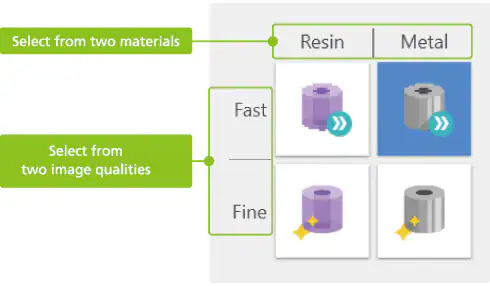
- Simplified scan mode selection
The system is equipped with a recommended scanning function that can configure all scanning conditions with a single mouse click. Optimal scanning conditions can be easily set by selecting the necessary combination of material type and image quality.
Support for safety and user protection
- System status monitor
In addition to displaying the device status, including the stage position during normal operation, the monitor provides detailed information about any errors that occur during abnormal operation and the location where the problem occurs, so the operator is immediately notified of any issue that may arise in the device.
The system status is continuously monitored, ranging from warnings that the sliding door is not closed, for example, to alarms indicating overheating of the X-ray generator and communication failures.
Specifications
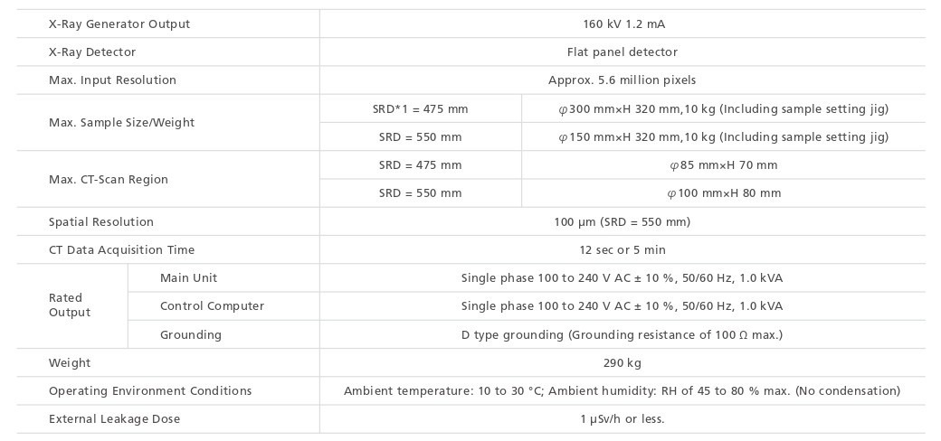
*1 SRD: Source to Rotation center Distance (SRD) is the distance from the X-ray source to the center of rotation of the sample.